Free sample lesson · Cell Membrane Exam Prep Essentials
Phospholipid Bilayer
How does a microscopic layer of oil keep a cell alive?
Lesson 1 of the Wondering course Cell Membrane Exam Prep Essentials
A Boundary Made of Oil
Every cell is wrapped in a boundary far too thin to see.
That boundary is built mostly from phospholipids. These are a type of lipid, the chemical family that includes fats and oils.
So the hook is literal. A microscopic layer of oily molecules separates the cell interior from its surroundings.
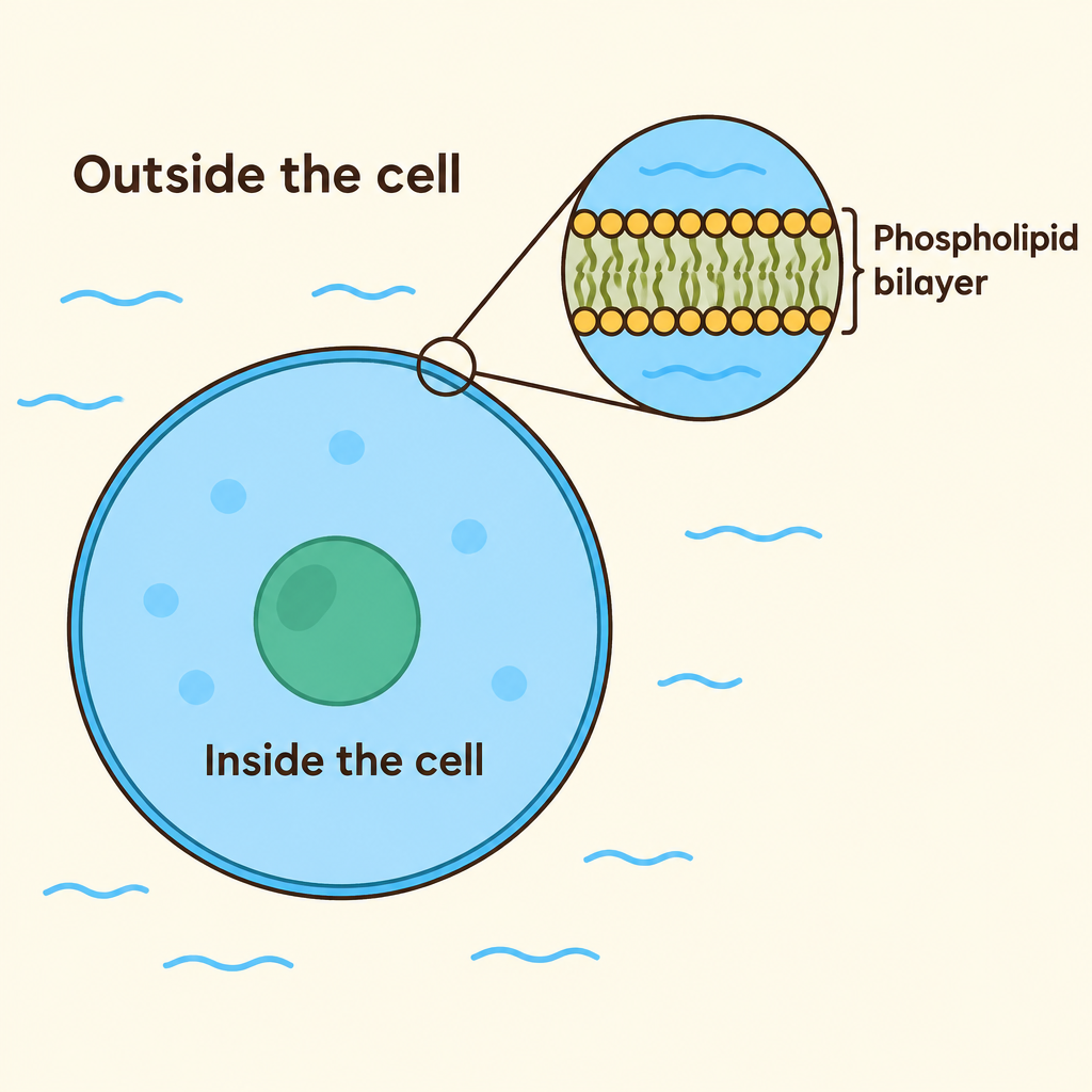
Notice how a very thin double layer forms the entire edge of the cell, separating the watery inside from the watery outside.
This structure is the phospholipid bilayer, a core feature of membrane structure. To understand it, start with one molecule.
One Molecule, Two Personalities
A phospholipid has two very different ends.
Biologists call this amphipathic. It means one molecule has both a water-loving part and a water-fearing part.
The head is hydrophilic, meaning it is attracted to water.
The tails are hydrophobic. They avoid water, much like oil avoids mixing into a glass of water.
- Head
- Tails
Their opposite behavior drives everything that follows.
Why Two Layers Form
Cells sit in watery surroundings, and their insides are watery too. So phospholipids meet water on both sides.
A double layer satisfies both ends at once. Heads point outward toward the water, and tails tuck inward, away from it.
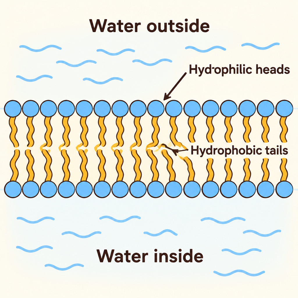
Follow the heads: both rows face water, while the tails from the two layers meet in the middle where no water reaches.
Nothing has to arrange this by hand. The chemistry of heads and tails pushes the molecules into this shape.
This tail-to-tail sandwich is the bilayer.
Reading the Membrane Zones
Picture a slice through the membrane from outside to inside. You cross three zones.
First, a row of hydrophilic heads touching the watery fluid outside the cell.
Next, the hydrophobic core, where tails from both layers meet. Water is kept out of this middle zone.
Last, a second row of heads facing the watery interior of the cell.
Exam tip: if a diagram shows tails touching water, it is wrong.
Flexible Yet Still a Barrier
The bilayer is a flexible double layer. Its molecules are held in place by how they respond to water, not bolted together like bricks.
That flexibility lets the membrane shift and bend.
Yet it still works as a boundary. It separates the cell interior from its surroundings while the cell interacts with its environment.
The key is that heads keep facing water and tails keep hiding, even as the layer moves.
From Chemistry to Survival
Now connect lipid chemistry to cell survival. Exam questions often test this exact link.
Because heads face water and tails hide inside, the bilayer forms a continuous boundary around the cell.
That boundary helps the cell maintain internal conditions, keeping its inside distinct from the outside.
Without it, the cell interior would simply mix with its surroundings. A cell cannot stay alive that way.
- Amphipathic molecule: Each phospholipid has a hydrophilic head and hydrophobic tails.
- Double layer: Heads face water on both sides while tails tuck inward.
- Continuous boundary: The bilayer separates the cell interior from its surroundings.
- Stable interior: The cell maintains its internal conditions.
- Cell survives: The inside does not simply mix with the outside.
Your Exam Takeaways
Here is what to bring into the exam.
Define it: phospholipids are amphipathic, with hydrophilic heads and hydrophobic tails.
Describe it: they form a flexible double layer. Heads face the watery inside and outside, and tails face inward.
Explain it: this arrangement separates the cell from its surroundings and helps it maintain internal conditions.
- Define
- Amphipathic phospholipids
- Hydrophilic heads, hydrophobic tails
- Describe
- Flexible double layer
- Heads out, tails in
- Explain
- Separates inside from outside
- Maintains internal conditions
Red flag on diagrams: tails should never touch water.
If you can say all three in plain words to a classmate, you are ready.
Check yourself
In a phospholipid, the fatty acid tails are _____
- hydrophobic
- hydrophilic
Answer: hydrophobic
Phospholipids are amphipathic: the phosphate-containing head is hydrophilic (water-loving), while the fatty acid tails are hydrophobic (water-fearing) and avoid contact with water.
In the phospholipid bilayer, the _____ face the watery fluid inside and outside the cell, while the _____ point inward toward each other, away from water.
Answer: In the phospholipid bilayer, the hydrophilic heads face the watery fluid inside and outside the cell, while the hydrophobic tails point inward toward each other, away from water.
Heads are attracted to water, so they line both surfaces that touch the watery cytoplasm and extracellular fluid. Tails avoid water, so they cluster in the middle of the bilayer.
Why do phospholipids placed in water arrange themselves into a double layer?
- Heads seek water and tails avoid it, so tails hide between two layers of heads
- The tails form strong covalent bonds with each other across the two layers
- The heads repel water, so they cluster together in the middle of the sheet
- The cell uses ATP to push each phospholipid into its correct position
Answer: Heads seek water and tails avoid it, so tails hide between two layers of heads
Because each phospholipid is amphipathic, the most stable arrangement in water exposes the hydrophilic heads to water on both sides while hiding the hydrophobic tails in the interior. No bonds between tails or energy input are required; it is driven by how each region interacts with water.
Keep going
This is lesson 1 of 11 in Cell Membrane Exam Prep Essentials. Next up:
- Fluid Mosaic
- Membrane Proteins
- Mechanisms of Membrane Protein Functions
- Simple Diffusion
- Osmosis
Wondering turns any topic into short lessons you can read, watch, or listen to, with practice questions and an AI tutor.